(63) The Clinical Utility of Spectral CT in Interventional Oncology: Innovations in Diagnosis and Intraoperative Guidance
Saturday, October 18, 2025
6:00 PM - 7:30 PM East Coast USA Time
Moaz Choudhary, MD – Interventional Radiologist, Department of Radiology, University of Texas Southwestern Medical Center; Patrick Sutphin, MD – Interventional Radiologist, Division of Vascular and Interventional Radiology, Massachusetts General Hospital; Sanjeeva Kalva, MD – Professor and Vice Chair of Image-Guided Interventions, Department of Radiology, University of Texas Southwestern Medical Center
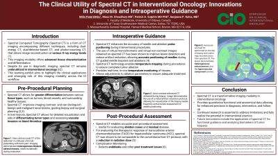
Purpose: Spectral Computed Tomography (Spectral CT) is a form of CT imaging encompassing different techniques, including dual-energy CT, dual-detector-based CT, and photon-counting CT, that allows image reconstruction at various X-ray energy levels. This imaging modality offers advanced tissue characterization and differentiation by acquiring images at multiple energy levels. Despite its use in diagnostic imaging, spectral CT remains underutilized in interventional oncology (IO). This learning exhibit aims to highlight the clinical applications and emerging role of this imaging modality across the IO workflow.
Material and Methods: This exhibit synthesizes recent literature and institutional case examples to illustrate the role of spectral CT in interventional oncology. Key applications include: 1. Pre-procedural planning: Improved differentiation of benign and malignant lesions using iodine maps, virtual non-contrast imaging, and extracellular volume quantification. 2. Intraoperative guidance: Real-time needle/probe positioning and lesion targeting using virtual monoenergetic images, thermal ablation thermometry. 3. Post-procedural assessment: Enhanced evaluation of ablation efficacy, detection of residual disease, and monitoring of complications such as endoleaks.
Results: Spectral CT improves diagnostic accuracy, interventional planning, and evaluation for treatment response and outcomes. Case examples demonstrate reduced contrast requirements in vascular imaging, accurate needle placement during biopsy and ablation, and improved detection of therapeutic response.
Conclusions: Spectral CT is a transformative imaging modality in interventional oncology. Its ability to provide quantitative functional and anatomical data allows for enhanced precision in diagnosis, intervention, and follow-up. Continued research is essential to address current limitations and to fully realize the potential of this technology in interventional practice. Such advancements will not only enhance its clinical utility but also contribute to broader adoption and lasting progress in the field of interventional oncology.