(34) A Quantitative Evaluation of a Calibration Phantom for CT-based Dosimetry following Yttrium-90 Radioembolization
Saturday, October 18, 2025
6:00 PM - 7:30 PM East Coast USA Time
Alasdair Syme, PhD – Medical Physicist, Dalhousie University; Aravind Arepally, MD – Chief Medical Officer, ABK Biomedical Inc.; Cheenu Kappadath, PhD – Medical Physicist, University of Texas MD Anderson Cancer Center
Purpose: A prerequisite for clinical CT-based dosimetry following yttrium-90 radioembolization (90Y-RE) is a comprehensive understanding of the relationship between Hounsfield units (HU) and radiopaque microsphere concentration. To this end, a calibration phantom was designed for clinical implementation. Microsphere detectability and the correlation between HU and microsphere concentration were evaluated. Phantom uniformity and reproducibility were also investigated.
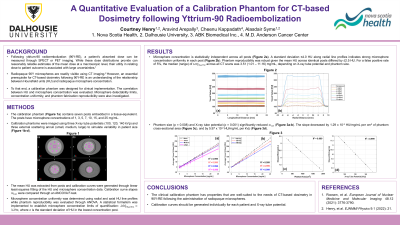
Material and Methods: A calibration phantom was designed to contain seven posts embedded in a tissue-equivalent resin. Each post contained uniformly distributed microspheres having concentrations between 1 and 25 mg/mL. The phantom was imaged with multiple CT scanners, a range of image acquisition parameters, and with an external scattering annuli to simulate variability in patient size. Microsphere concentration uniformity was determined using radial and axial HU line profiles while phantom reproducibility was evaluated using ANOVA. A statistical formalism was implemented to establish microsphere concentration limits of detection (LOD). Reconstructed CT images were segmented using MIM software. The mean HU was extracted from post structures and calibration curves were generated through linear least-squares fitting of the HU and microsphere concentration data. Calibration curve slopes were compared through an ANCOVA F-test.
Results: Line profile variability of ≤ 4.0 HU indicated strong microsphere concentration uniformity. Phantom reproducibility was robust given the mean HU across identical posts differed by ≤ 2.5 HU. The median (range) of LOD across all CT scans was 5.18 (1.12 – 13.42) mg/mL for a false positive rate of 5%. Changes in image noise produced non-statistically significant differences in the calibration curve slope. Conversely, increased phantom thickness (p = 0.008) and increased tube potential (p = 0.001) significantly reduced the calibration curve slope.
Conclusions: The calibration phantom has properties that are well-suited to the needs of CT-based dosimetry in 90Y-RE following the administration of radiopaque microspheres. Calibration curves should be generated individually for each patient and CT tube potential.